Rethink Imaging Podcast Transcript
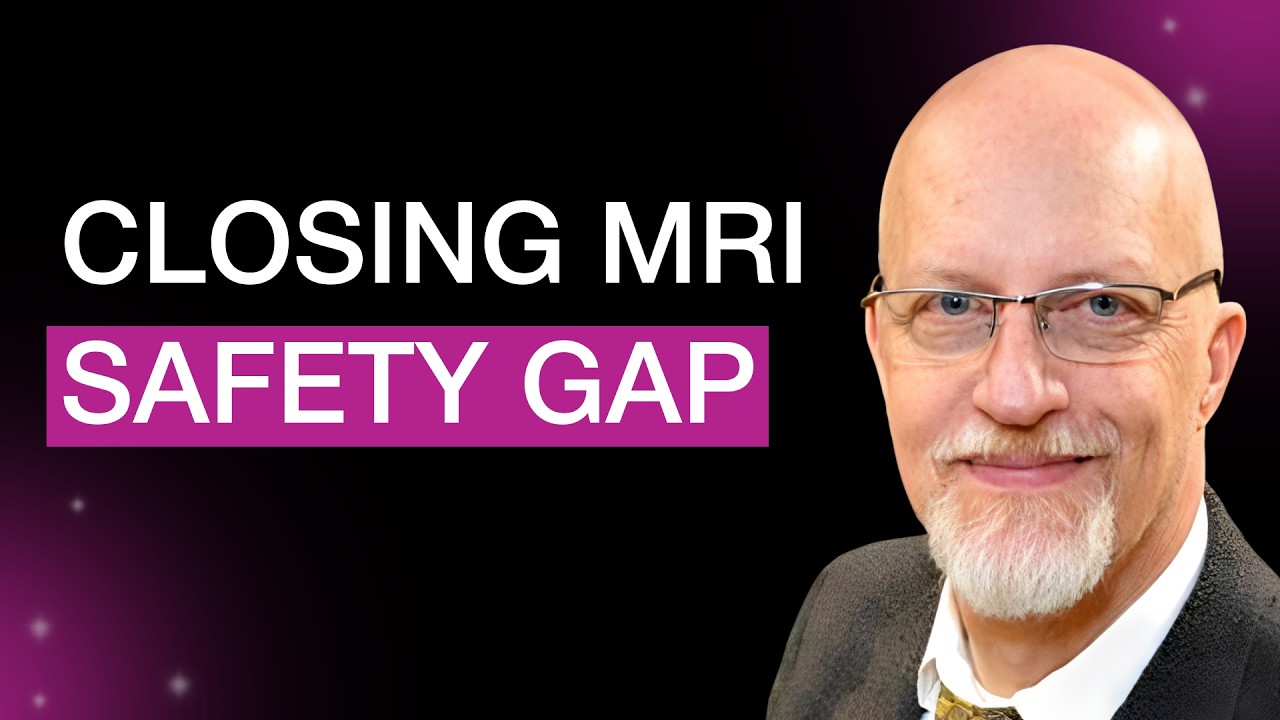
Guest: Tobias Gilk
Host: Chris St. John
[00:00:28] Chris St. John: We’re here because we’re curious about medical imaging or have a hunger to learn more. What can we learn from each other, and how should we prepare to tackle the challenges ahead? I’m Chris St. John, and join me today as we Rethink Imaging, a podcast by Imalogix. Welcome to the show. Welcome back to Rethink Imaging. We’re joined today by Tobias Gilk, it’s so great to have you today.
[00:00:54] Tobias Gilk: Thank you so much. I’m so thrilled to be here with you.
[00:00:57] Chris St. John: Before we start getting into all of the nuance of MRI safety, I want to talk a little bit about you and let our listeners get to know you as well. Do you mind talking me through your pathway through architecture and into MRI a little bit?
[00:01:19] Tobias Gilk: There were a whole bunch of unplanned left-hand turns in my career. When I was doing my undergraduate, I was actually working at the theater where a bunch of traveling shows came through the university. A concert series would be Monday night, Tuesday night would be a student film, Wednesday night would be a lecture, Thursday night would be a dance recital, and Friday night would be the film series or something like that. We were constantly turning over this theater building. For somebody working in that space, you become intimately aware of what about the design of that building doesn’t really work or doesn’t really support the intended function.
I actually went to architecture school because I wanted to design theaters—forget this theater design and production stuff. I probably should have done a little bit of homework before then, realizing that in the United States, we don’t really build live theaters anymore, so career opportunities for an architect were limited. I pretty much shelved that as some sort of grand plan, went to architecture school, got my master’s in architecture, came out, and worked for a small firm in a small college town. That firm had done essentially zero healthcare before I got there, but they had been bugging the county hospital’s director of design and construction, saying, “Hey, give us a shot. Give us a small project. We’ll prove our worth.”
Coincidentally, right when I got there, the county hospital’s director of design and construction—I guess they wore him down—said, “Okay, fine. I’ll give you guys a little project to prove yourself.” The owners of the firm looked around and realized all of their architects were already assigned to other projects and they couldn’t pull one of them off. So, they gave them the new kid. They essentially gifted me to the hospital and said, “Whatever you need him to do, he will do.” So, I converted a large storeroom into an office for the director of surgical services.
That went fairly well because the next thing I knew, I was doing the very exciting design work to install a pneumatic tube station in the hospital’s ICU. I guess that went well and I was making friends over at the hospital. They wound up giving us this ridiculously fast-paced MRI project at the end of the year. I guess they felt like they could work with me and work with the firm. I was less than six months out of grad school, and I was managing the design and installation of a new MRI suite.
One of the things I didn’t realize at the time was that architects develop a set of skills where they figure out what they don’t need to know about their client’s business. Not only did I not have those skills, I didn’t even recognize that that was a skill set an architect developed. So when they told me I was doing an MRI project, I felt I had to learn everything there is to know about MRI. I just threw myself into the deep end trying to figure everything out about this technology, and I was hooked. I’ve always been a science-geeky kind of person, and MRIs are the closest thing to magic that human beings have ever created. So, I was hooked on MRIs.
But this was 1997, before there was any real awareness of MRI safety. Fast forward to 2002: the same hospital was adding a third magnet—the one we did before was their second magnet—because they kept growing this service, and they brought me back again. In 2002, the ACR published what was then the first white paper on MR safety, the very first document in the United States that says, “Here’s how we should do MRI more safely.” It was published in response to a horrific accident that happened the year prior. The head MRI technologist handed this to me and said, “Hey, whatever it says, do.”
Well, I had a panic attack moment because nobody told me I had to pay attention to any of the safety requirements when I did the one five years prior. What all did I miss? I didn’t look at the state requirements for all of this. After a day or two of panic and frantically looking up what the safety requirements were for MRI suites, I discovered there are none. From that moment, I said, “Well, this is dumb. We should have some sort of minimum safety standards from an architectural design and construction standpoint.”
That was really the snowball being pushed down the very top of the mountain, and it began to collect momentum en masse. That beginning of focusing on the bricks-and-mortar part of MRI safety just metastasized over the years, and I became interested in all aspects of MRI safety.
[00:06:49] Chris St. John: It’s so funny. I identify with this story a lot. I took a lot of architecture classes in college myself, actually, and my senior thesis—while it was an installation—I called a choreography of a space, about how we view art in a gallery setting, how we move in a gallery setting. What you were talking about in terms of designing a better theater, I think that all translates very clearly into the design of an MRI space: thinking about intention, thinking about best practices, thinking about safety. It all translates. It feels like a very smooth transition to me, in fact.
[00:07:33] Tobias Gilk: If you stop and ask somebody what a live production theater and an MRI suite have in common, you probably could not pick two more different spaces. But I look at it very much the way you just described, and I’m doing exactly the thing that was my intention when I decided to go to architecture school. I just changed the building that I’m doing this for. I’m no longer doing it for theater buildings; I’m doing it for radiology departments.
[00:08:04] Chris St. John: Here you are, discovering that there are no safety requirements for these MRI suites. Let’s bring in some of your public discourse. You have said—I think of it as your tagline—that MRI is not the safe modality. When compared to CT or X-ray, I think a lot of people’s gut instinct would say, “Of course it is.” Would you mind expanding on that and explaining what you mean?
[00:08:43] Tobias Gilk: I think in radiology, we are victims of our own PR relative to MRI and our messages about MRI safety. I think everybody who has a passing familiarity with radiology has heard and probably internalized to some level that MRI is the safe imaging option. We’ve all heard that. We all understand that ionizing radiation has some inherent risks, and if we could choose an imaging option that doesn’t have the same inherent risks, that seems to be the logical choice. What that bumper-sticker slogan for MRI doesn’t convey is that, yes, MRI doesn’t have the ionizing radiation risks, but it has its own unique, peculiar risks just within MRI.
It appears to me that the conversation when the MRI first came upon the scene was something along the lines of, “Hi, we’re MRI. We’d like to get official approval to be able to sell our equipment.” And the regulatory world was like, “Yeah, we’re really worried about ionizing radiation. Tell us how much ionizing radiation the patient gets with an exam in your new machine.” “Well, none.” “Oh, okay. In which case, you’re approved.” There was never the follow-on conversation of, “Well, if you don’t have those sets of risks, what risks do you have?”
It’s almost like somebody looked around and said, “Oh my goodness, 10,000 people die in car crashes in the United States every year. If we want to eliminate deaths by automobile accident, the thing that we should do is outlaw cars and just have everybody parachute to work. Jump out of a plane with a parachute on, and car crash deaths will go to zero.” While that’s true, that ignores the fact that there are different, unique risks associated with skydiving. Now, I’m not trying to equate the greater risk of skydiving with some greater risk in MRI—that’s not the case—but we need to acknowledge that there are different sorts of risks and we need to respond to the different sorts of risks in different ways.
In, I think it was 2006, there was a toddler who got a CT scan at a facility in Mad River, California. And the CT tech—little two-year-old squirmy kid was not holding still, not cooperating for the head CT—so the CT tech just hit the scan button over and over again, more than 100 times. This incident was so egregious in the world of radiation safety that it was all over the news. The pictures of the kid, you could see what were essentially sunburns, radiation burns, around the circumference of the kid’s entire head, and that was the worst accident. There was identified chromosomal damage within this kid. I do not mean to make light of this or minimize that in any way, shape, or form.
That was 2006, and I have not heard of a single other radiation exposure incident that has made the press since 2006, so we’re 20 years on. I haven’t heard of a single one that has made the press. We’re really good at hiding our dirty laundry, so I don’t mean to suggest that nothing has happened since then. But in that intervening time period in MRI, we saw a man with an aneurysm clip die in a hospital in Southern California. We saw, a few years ago, a nurse get crushed by an ICU bed against an MRI scanner. Last year in the state of New York, we saw a man get killed by the chain around his neck. Same time frame last year, there was a pacemaker ICD patient who died in an outpatient imaging center in Tennessee. A couple of years before, there was a patient who was hit by a flying wheelchair and knocked off of the MRI table as the wheelchair went flying at the magnet. There was another patient who had an esophageal temperature probe—essentially, you swallow this probe and it monitors your body temperature from inside you, but there’s a wire that comes out of the mouth. This patient underwent an MRI, and the wire heated up, giving the patient burns in their throat, tongue, mouth, and lips.
So we have all of these incidents where we have very significant safety issues in MRI, and yet we run around and recite the bumper-sticker slogan, “MRI is the safe imaging option,” without any meaningful protections for patients or even healthcare workers who work in these environments.
[00:14:29] Chris St. John: I’d love to expand on those examples. It’s my understanding that you and Dr. Emmanuel Kanal were doing some work with the FDA where you were exploring the different dangers of MRI, and I believe that y’all documented something like 95 percent of MRI injuries fall into those categories: burns, projectiles, hearing damage.
[00:14:57] Tobias Gilk: Ninety-five percent of the ones that are cataloged as MRI accidents. If an MRI causes a pacemaker or an insulin pump to malfunction and things go badly, those accidents—even though the MRI was the reason for the malfunction—just get cataloged today as malfunctions of a pacemaker or malfunctions of an insulin pump. So we actually have no good way to track interactions between the MRI scanner and implants and devices.
Yes, of the injury accidents that get cataloged under “this was an MRI incident,” 95 percent of the injury accidents are burns, projectiles, and hearing damage. And we have some really remarkable best practice standards that can interrupt the way that those accidents happen, and yet nobody requires them. No states, no accreditation organizations. CMS doesn’t require it as conditions of getting paid for Medicare or Medicaid patients. They just don’t exist.
[00:16:09] Chris St. John: And why do you think that is?
[00:16:12] Tobias Gilk: I think it’s a combination of reasons. I think it’s partially inertia. If you go from state to state, and between the various states and the federal government, and you’re looking at the radiation safety rules, they are remarkably consistent across all of the different states and between the states and the feds. Did everybody come up with their own set of rules and they just happen to match almost perfectly across all 50 states? No. What happened was, when we became concerned about ionizing radiation exposure in diagnostic imaging, human beings are lazy. We went looking for where there is a standard that we can copy or adapt. And what they found was that the U.S. Department of Energy developed a workplace safety standard for folks working on the Manhattan Project.
They were like, “The Department of Energy did our homework for us. We’re going to copy and paste the DOE workplace safety standards, and these are going to become our state’s ionizing radiation standards.” And that copy and paste essentially spread like wildfire. That’s why ionizing radiation standards are so uniform, in part because nobody had to do any work other than copying and pasting them.
Fast forward to MRI, there were no predicate safety standards about exposure to high-strength magnetic fields. There were no predicate safety standards on oscillating or pulsed electromagnetic fields and the safety risks. So we’re faced with the challenge of the blank canvas. Where do we start? What’s appropriate? Who has done work before to tell us if we’re going in the right direction or the wrong direction? I think the intimidation factor of not being exactly sure what it is that we’re doing, combined with regulatory inertia—who wants more regulation? Everybody’s like, “Look, until people start fussing about this, we’re going to let sleeping dogs lie.”
Now, the consequence of that has been there were all of these preventable accidents—accidents where we know how the accident happens, accidents where we know what are effective interventions that would stop the chain of causation. And yet, from a regulatory and accreditation standpoint, we all collectively throw up our hands and say, “Boy, man, I wish there was something we could do,” ignoring the fact that there is, in fact, a lot we can do.
[00:19:09] Chris St. John: I want to come back to that. Something you said when you were talking about the copying and pasting of safety standards triggered something for me. When we spoke previously, one of the things you mentioned was that a lot of MRI workflows were inherited from CT and never really updated. It’s almost this additional copy-paste over. I’m curious, with that workflow copy-and-paste over, can you walk us through what that looks like in practice a little bit?
[00:19:40] Tobias Gilk: Sure. For folks who haven’t seen or aren’t conjuring in their minds a picture of a CT scanner and a picture of an MRI scanner—and for that matter, the picture of a PET scanner—these are almost identical-appearing pieces of equipment. We have a big donut-shaped scanner, and then there’s a table that sticks out of it like a tongue. You get on the table, the table slides you into the middle of the donut, and you get a scan.
CT scanners, MRI scanners, and PET scanners look remarkably similar. CT scanners were the first ones on the scene, and we realized we want to make sure that the operator of the CT scanner is not being exposed to the radiation. We’re going to build a separate control room for them. And we’ve got all of this equipment that doesn’t have to be in the room with the CT scanner but is necessary, so we’ll build a little equipment room associated with this. Then we’ll get our patients ready and have a little area for them to rest so that we can grab the next patient and bring them in. Some of the patients will probably need to change at least partially out of their clothes, making sure there are no zippers, snaps, or buttons in the field of view so that we can get good, clean CT pictures.
For those folks who are familiar with MRI, they’re scratching their head right now going, “Why is he describing an MRI workflow?” Because that is essentially what happened when MRI came upon the scene after CT. We just looked at what we had invented before, and we said, “You know what? That’s probably going to work.” Now, at the time, a CT scan was probably 20 minutes, and an MRI study was probably going to be an hour and a half. So we didn’t even really need to worry too much about patient preparation and sub waiting because MRI was such a slow process. There would be lots of time to get people ready and that kind of thing.
Well, MRI has sped up dramatically, and we’re beginning to see how the workflows and the demands of getting people ready are very different in the MRI space. In CT, we can almost just take you off the street, make sure that there’s nothing you’re wearing or carrying—jewelry or whatever—that’s going to interfere with the image, and we can walk you right into the CT room, throw you on the table, scan you, and walk you out of the building in less time than it will take to get that MRI patient to fill out a screening form. We need to identify: Do you have an aneurysm clip? Do you have a pacemaker? Did you have a pacemaker where they took most of it out but left fragments of the leads in? We need to identify all of these things, and if any one of those answers is, “Yes, I do have that in me,” well, great. Now we have to go and look up your medical records. Now we have to figure out exactly what that thing is within you.
This is no exaggeration: it would not surprise me in certain circumstances, if we get an easy CT patient and a difficult MR patient, if we could scan six CT patients in the time that it takes us to do the clearance activities for one MRI patient. So the clearance and the preparation for MRI takes a significant chunk of time.
Now the scanners are accelerating and are able to produce these exams in less time, so we need to focus more and more attention on patient screening and patient preparation. Are we gowning the patients? Where is that happening? How are we doing the sub ways? We are seeing, through the acceleration of MRI scans and greater emphasis and greater complication in patient screening and preparation, an emerging conflict within MRI. It really requires, if we’re going to tackle this smartly, that we rethink the entire workflow around MRI. It ought not be a copy and paste of CT because the demands are very different.
[00:24:12] Chris St. John: Are you comfortable sharing some of your thoughts and opinions on like what an idealized MR workflow could look like?
[00:24:22] Tobias Gilk: First off, it’s going to be very different if we’re talking about an outpatient imaging center versus a hospital, because outpatient imaging centers generally get to cherry-pick the less complicated patients. But if we’re looking at a hospital setting, one of the things that really ought to happen is we ought to be pushing more of the patient screening and patient preparation upstream.
It has been the practice that, other than being told when to show up for your MRI, that was probably the one and only thing you were told beforehand. You show up there, they describe what the exam is going to be, they hand you the screening form, and we’re doing all of this checking you out in the 30 minutes prior to your scheduled exam, or however that works. We need to be looking, because of the complexity of managing these patients, at how we institutionalize patient review, patient prep, and those kinds of activities so that when the patient shows up, we already know what’s in them, on them, or with them. We’ve already had an opportunity to look that up. If there were questions or concerns, we’ve had the opportunity to resolve those. When the patient shows up, none of that preparatory work other than getting them changed is really necessary. We want to diminish and reduce the amount of time at the point of care for all of that preparation and screening activity work.
So now we have patients there. If our MRI studies are averaging 20 or 25 minutes apiece, we need to be expecting how we are going to be efficiently moving the patient who’s in there right now out, how we’re moving the next patient in, and how we’re completing whatever preparation works are necessary for patient number three. Nobody likes feeling like they’re on an assembly line, but from a workflow standpoint, we need to be thinking of it like an assembly line, like a machine. And there are lots of things you can do to make people’s experience of that assembly line or machine feel much better. Just ask Disney World. How long do we stand in line for one of their rides? Every twist and turn in the line, you get to see something new.
So we can engineer not only the workflow, but we can engineer the patient experience so that it doesn’t suck. We need to be rethinking all of these things in terms of how we are going to maximize the available productivity of these machines that are faster and better. How do we get our money’s worth out of those machines and, at the same time, do that in a way where we’re not pushing people through the preparatory process for MRI?
Rethink Imaging is brought to you by Imalogix. Here, you’ll find engaging interviews with thought leaders, experts, and patients sharing stories that showcase the transformative power of medical imaging. To discover how Imalogix is rethinking imaging in healthcare, visit imalogix.com. Be sure to subscribe to Rethink Imaging on Apple Podcasts, Spotify, or wherever you listen. And from all of us here at Imalogix, thanks for tuning in.